Label the structures of a synovial joint, the most common type of joint in the human body, and delve into the intricate world of joint anatomy. This guide will provide a comprehensive overview of the various components that make up a synovial joint, their functions, and their importance in maintaining joint health and mobility.
From the smooth articular surfaces to the stabilizing ligaments, each structure plays a vital role in ensuring joint stability, facilitating movement, and absorbing shock. Understanding the anatomy of a synovial joint is crucial for healthcare professionals, fitness enthusiasts, and anyone interested in maintaining optimal joint function.
Joint Structure and Function

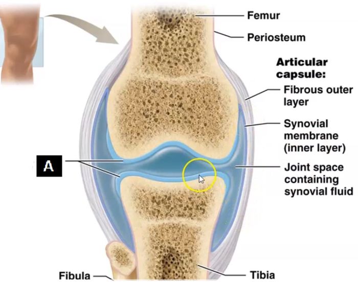
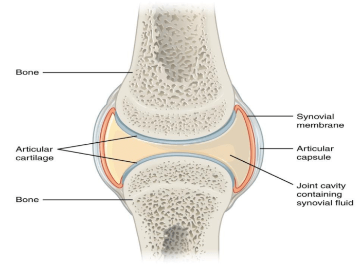
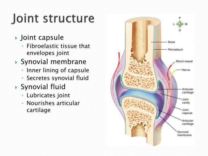
Synovial joints are the most common type of joint in the human body. They are characterized by the presence of a synovial membrane, which lines the joint cavity and produces synovial fluid. Synovial fluid is a viscous fluid that lubricates the joint and provides nutrients to the articular cartilage.
The articular surfaces of synovial joints are covered with a layer of hyaline cartilage. Hyaline cartilage is a smooth, glassy cartilage that provides a low-friction surface for movement. The joint cavity is filled with synovial fluid, which provides lubrication and nutrients to the joint.
Functions of Synovial Joints
- Movement: Synovial joints allow for a wide range of movements, including flexion, extension, rotation, and abduction.
- Stability: Synovial joints are stabilized by a network of ligaments and muscles.
- Shock absorption: The synovial fluid in synovial joints helps to absorb shock and protect the joint from damage.
Joint Cartilage

There are three types of cartilage found in synovial joints: hyaline cartilage, fibrocartilage, and elastic cartilage.
Hyaline Cartilage
Hyaline cartilage is the most common type of cartilage in the body. It is found on the articular surfaces of synovial joints and provides a smooth, low-friction surface for movement.
Fibrocartilage
Fibrocartilage is a strong, flexible cartilage that is found in intervertebral discs and menisci. It provides cushioning and support to these structures.
Elastic Cartilage
Elastic cartilage is a flexible cartilage that is found in the ear and epiglottis. It allows these structures to bend and stretch without breaking.
Synovial Fluid: Label The Structures Of A Synovial Joint
Synovial fluid is a viscous fluid that fills the joint cavity. It is produced by the synovial membrane and provides lubrication and nutrients to the joint.
Synovial fluid is composed of water, proteins, and hyaluronic acid. Hyaluronic acid is a thick, gel-like substance that gives synovial fluid its viscous properties.
Functions of Synovial Fluid, Label the structures of a synovial joint
- Lubrication: Synovial fluid lubricates the articular surfaces of synovial joints, reducing friction and wear and tear.
- Nutrition: Synovial fluid provides nutrients to the articular cartilage, which is avascular.
- Shock absorption: Synovial fluid helps to absorb shock and protect the joint from damage.
Joint Ligaments

Ligaments are tough, fibrous bands of tissue that connect bones to each other. They help to stabilize synovial joints and prevent them from dislocating.
The major ligaments associated with synovial joints include:
- Anterior cruciate ligament (ACL)
- Posterior cruciate ligament (PCL)
- Medial collateral ligament (MCL)
- Lateral collateral ligament (LCL)
Functions of Joint Ligaments
- Stability: Ligaments help to stabilize synovial joints and prevent them from dislocating.
- Proprioception: Ligaments contain proprioceptive nerve endings that provide the brain with information about the position of the joint.
Joint Muscles

Muscles that cross synovial joints contribute to their movement and stability.
The major muscles associated with synovial joints include:
- Quadriceps femoris
- Hamstrings
- Gluteus maximus
- Deltoids
Functions of Joint Muscles
- Movement: Muscles that cross synovial joints contribute to their movement.
- Stability: Muscles that cross synovial joints help to stabilize them and prevent them from dislocating.
- Proprioception: Muscles that cross synovial joints contain proprioceptive nerve endings that provide the brain with information about the position of the joint.
Question & Answer Hub
What is the function of synovial fluid?
Synovial fluid nourishes and lubricates the joint, reducing friction during movement.
Which ligament is responsible for preventing excessive forward movement of the tibia?
The anterior cruciate ligament (ACL) prevents excessive forward movement of the tibia.